The ESP (Erector Spinae Plane) Block
Featured Free Videos
•
18m
The video was created by Dr. Vicente Roques, an anesthesiologist at el Hospital Universitario Virgen de la Arrixaca in Murcia, Spain, with contributions from Dr. Mauricio Forero and Dr. Ki Jinn Chin. They summarize the current (2017) understanding of the ESP block including anatomy, possible mechanisms, technique, and clinical pearls.
00:29 Muscle layers
00:40 Spinal / intercostal nerves
01:26 Muscles of the back
02:01 The intertransverse connective tissue complex
02:09 Conceptual view of ESP and the paravertebral space
04:31 HOW DOES THE ESP BLOCK WORK?
05:19 1-The costotransverse foramen
05:56 2-The intertransverse connective tissue complex
06:36 Conceptual view of the ESP block
06:59 SONOANATOMY OF THE ESP BLOCK
07:11 The T5 transverse view
07:33 The T5 sagittal paramedian view
08:53 THE ESP BLOCK APPROACH
10:11 HOW TO PERFORM AN ESP BLOCK
10:18 Before starting
10:41 Patient position
11:25 T5 transverse scanning in real-time
12:26 T5 sagittal paramedian scanning medial-to-lateral in real-time
13:31 T5 sagittal paramedian scanning lateral-to-medial in real-time
14:38 In-plane approach to the ESP block in real-time
16:34 INDICATIONS FOR THE ESP BLOCK
17:08 KEY POINTS 17:39 TIPS
Up Next in Featured Free Videos
-
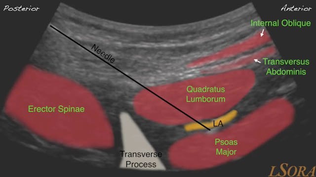
LSORA Quadratus Lumborum Block
This video tutorial was created by Dr Parthipan Jegendirabose (PJ) - Consultant Anaesthetist at Colchester General Hospital, UK, and Dr Amit Pawa, and Dr Mark Ibrahim who are both Consultant Anaesthetists at Guy’s & St Thomas’ NHS Foundation Trust in London, UK. Dr Amit Pawa is also the Regional ...
-
Blockjocks VIP Trailer
This video highlights the benefits of a subscription to the BLOCKJOCKS Virtual Interactive Preceptorship (VIP), the premium subscription available on blockjocks.com. To sign up for a one week FREE trial click here: http://www.blockjocks.com/buy/subscription
-
FREE Cadaver Demo- Saphenous Nerve & ...
In this FREE BLOCKJOCKS BASIC cadaver demonstration video Dr. Brianne Owens and Dr. Greg Hickman demonstrate the separate compartmentalization of the saphenous nerve and the nerve to vastus medialis (NVM) at the mid-thigh. This finding mirrors their clinical experience during ultrasound-guided ad...


